
Scanning electron microscope (SEM) Definition, Images, Uses, Advantages, & Facts Britannica
Electron microscope (EM) uses high-energy electron beam as probe instead of visible light. The electrons have shorter wavelength and provides very high-resolution capacity (0.1 nm) and 500,000 times magnification power. It is also easy to manipulate the electron beams. Instead of glass as lens, the electron microscope uses electromagnetic coil.
5 Science Facts Behind Astonishing Electron Microscope Images Rs' Science
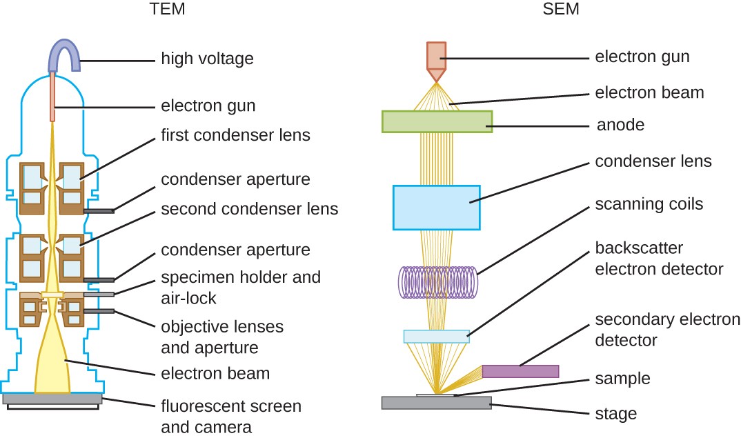
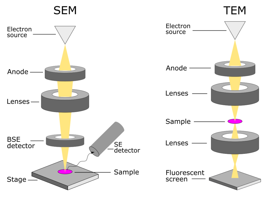
The transmission electron microscope (TEM) can image specimens up to 1 micrometre in thickness. High-voltage electron microscopes are similar to TEMs but work at much higher voltages. The scanning electron microscope (SEM), in which a beam of electrons is scanned over the surface of a solid object, is used to build up an image of the details of the surface structure.
"Electron Microscopes" Electron Microscopes (EMs) are scientific instruments that use a ray of
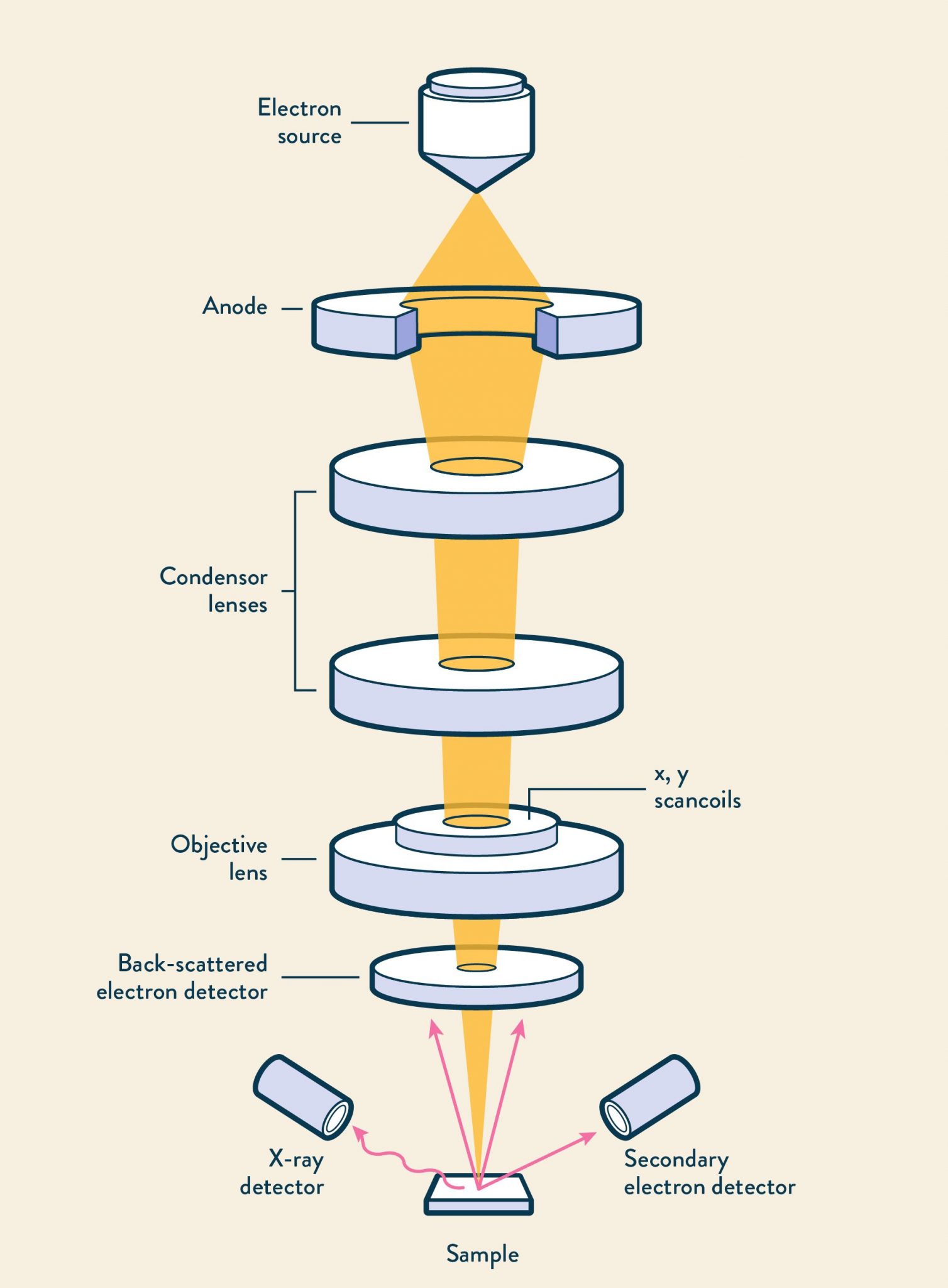
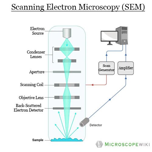
What is Scanning Electron Microscopy (SEM) A typical SEM instrument, showing the electron column, sample chamber, EDS detector, electronics console, and visual display monitors. The scanning electron microscope (SEM) uses a focused beam of high-energy electrons to generate a variety of signals at the surface of solid specimens.
Schematic flow diagram of a Scanning Electron Microscope. Electron... Download Scientific Diagram
Scanning Electron Microscopes (SEM). The transmitted electrons carry information about the structure of the specimen. The magnified image is formed by the objective lens system and is viewed by being projected onto a fluorescent viewing screen or a digital camera. Modern high-resolution TEM microscopes can reach magnifications well above.
Microscope Drawing Template at GetDrawings Free download
tissues the scanning electron microscope (SEM) has a large depth of field so can be used to examine the surface structure of specimens TEMs have a maximum magnification of around ×1,000,000,.
The components of a transmission electron microscope (TEM Stock Photo, Royalty Free Image
TEM: An Overview. Transmission electron microscopy (TEM) is a form of microscopy which in which a beam of electrons transmits through an extremely thin specimen, and then interacts with the specimen when passing through it. The formation of images in a TEM can be explained by an optical electron beam diagram in Figure 8.2.1.

SEM vs. TEM Electron Microscopy • Microbe Online
An electron microscope is a microscope that uses a beam of accelerated electrons as a source of illumination. It is a special type of microscope having a high resolution of images, able to magnify objects in nanometres, which are formed by controlled use of electrons in a vacuum captured on a phosphorescent screen.
Telescope vs Microscope What's the Difference? Optics Mag
Electron microscopes are used for detailed investigation of the ultrastructure of a wide range of biological and inorganic specimens including microorganisms, cells, large molecules, biopsy samples, metals, and crystals. German physicist Ernst Ruska invented electron microscope in 1931. Table of Contents Components of an Electron Microscope
Simple Microscope Drawing at GetDrawings Free download
Simple Microscope Diagram Compound Microscope Electron Microscope Stereo Microscope Scanning Probe Microscope Frequently Asked Questions - FAQs What Are the Different Types of Microscopes? There are different types of microscopes and each of these has different purposes of use.
Electron Microscope Principle, Types, Applications Microbe Online
2. Scanning Electron Microscope (SEM): An SEM creates magnified images of the specimen by probing along a rectangular area of the specimen with a focused electron beam. This process is called the raster scanning.
Instruments of Microscopy Microbiology Course Hero
The electron microscope uses a beam of electrons and their wave-like characteristics to magnify an object's image, unlike the optical microscope that uses visible light to magnify images. Conventional optical microscopes can magnify between 40 to 2000 times, but recently what are known as "super-resolution" light microscopes have been developed.

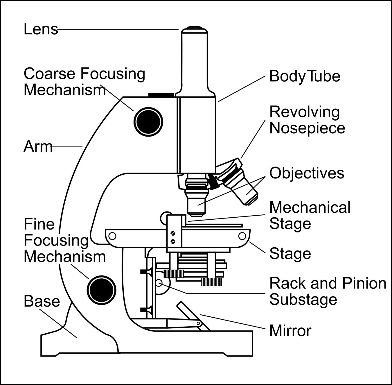
Microscope Diagram to Print 101 Diagrams
An electron microscope is a microscope that uses a beam of electrons as a source of illumination. They use electron optics that are analogous to the glass lenses of an optical light microscope to control the electron beam, for instance focusing them to produce magnified images or electron diffraction patterns.

Simple Microscope Drawing at GetDrawings Free download
In an electron microscope, a stream of electrons takes the place of a beam of light. An electron has an equivalent wavelength of just over 1 nanometer, which allows us to see things smaller even than light itself (smaller than the wavelength of light's photons).
Electron Microscopy AnaPath
What is electron microscopy? What is the diference between scanning electron microscopy (SEM) and transmission electron microscopy (TEM)? How can you choose a microscope that best fits your research process? CHAPTER 2 Challenges in microscopic analysis
Scanning Electron Microscope Block Diagram
Scanning Electron Microscope (SEM) is a type of electron microscope that scans surfaces of microorganisms that uses a beam of electrons moving at low energy to focus and scan specimens.
Electron Microscope Principle, Uses, Types and Images (Labeled Diagram), Price
An electron microscope (EM) uses a high energy electron beam aa s probe instead of visible light. The electrons have a shorter wavelength and provide a very high-resolution capacity (0.1 nm) and 500,000 times magnification power. It is also easy to manipulate the electron beams. Instead of glass as a lens, the electron microscope uses an.